RAYSCAN S
All-In-One Scanner For Every Clinical Specialty
With a large, adjustable 20×20 field of view for evaluating pathology, airway assessments, TMJ evaluations, surgical planning, and complex restorative cases, the RAYSCAN S integrates pan, CBCT, a CT impression (intraoral data) scan, and an optional ceph into a perfect imaging machine. The revolutionary visible light guide makes it easy for users to always take the perfect scan, eliminating the need for retakes. In addition, the S increases production and spares patients the uncomfortable experience of intraoral sensors by extracting an FMX from a single pano image.
Light Guide
Free FOV
20×20 FOV
70μm Voxel
Fast Scan
Low Dose
See where you focus
Light-Guided Free FOV
Dose Reduction
CBCT
Patient Safe and Easy Operation
All-In-One Scanner For Every Clinical Specialty
RAYSCAN S provides accurate diagnostics for treatment that can be applied to all forms of digital dentistry.
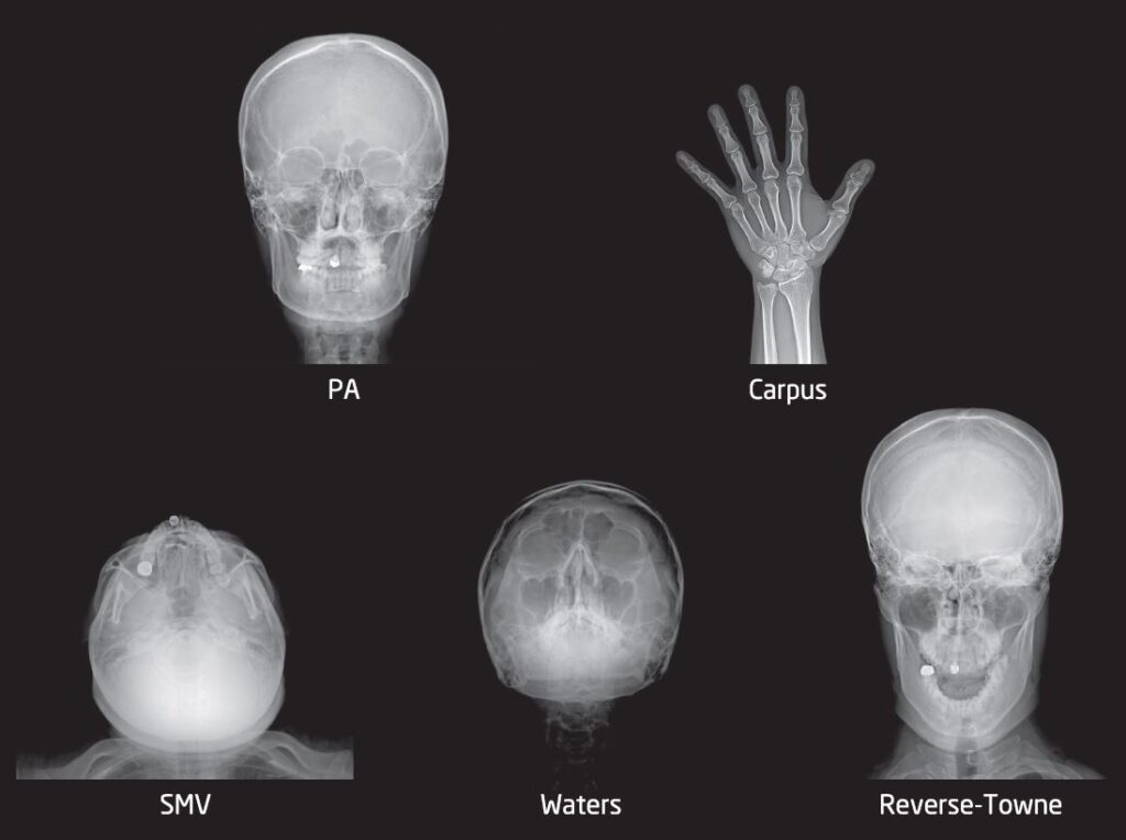
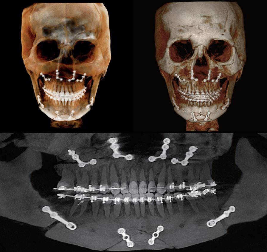
Pre-operative and post-operative assessment.
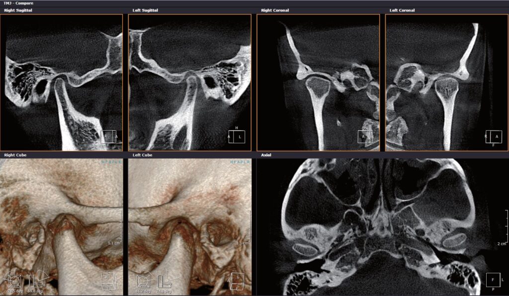
Capture both left and right TMJ with a single capture; the 200 μm TMJ images aid to diagnosis a variety of TMD.
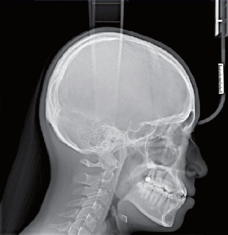
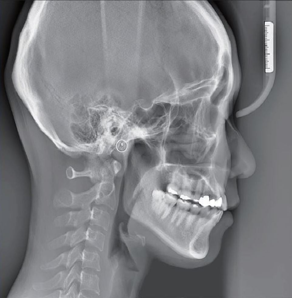
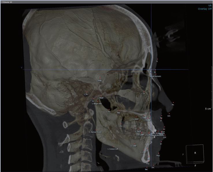
3D radiographic analysis supports accurate diagnosis & optimal treatment objectives.
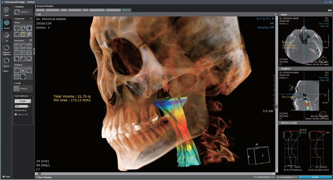
Excellent visualization of static airway morphology.
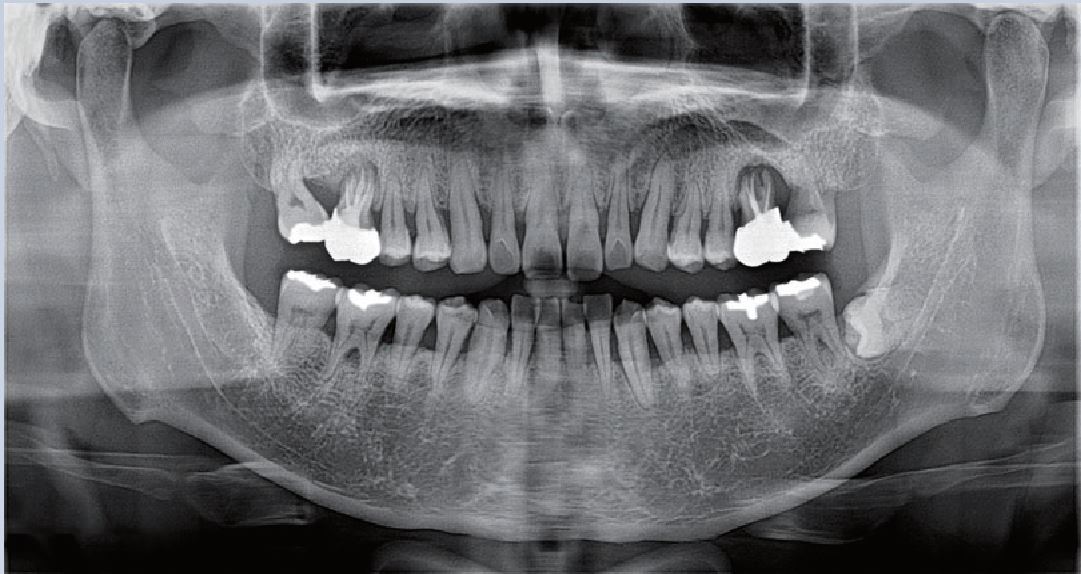
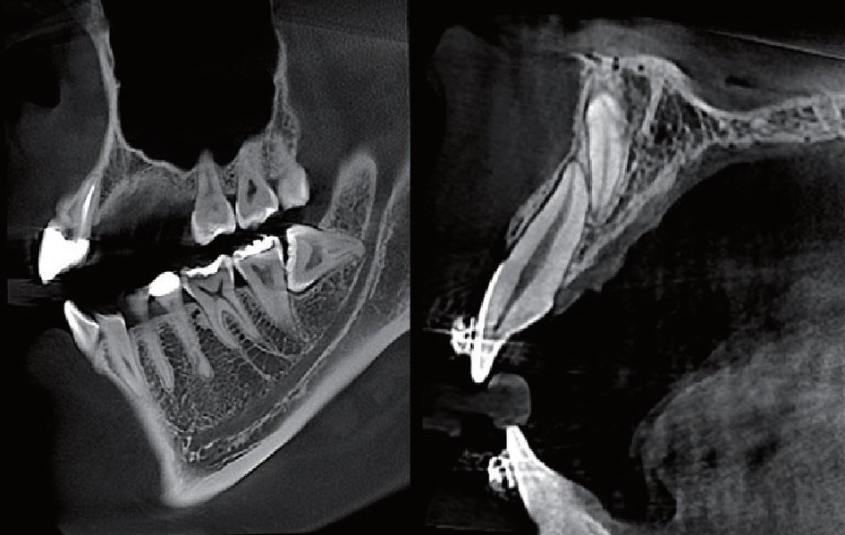
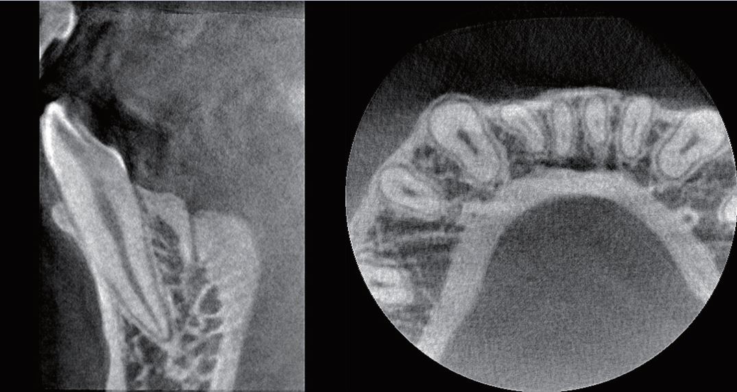
Determine exact location of impacted teeth and assess for root resorption on adjacent teeth.
Scan and reconstruct at 70μm with a focused field of view for precise endodontic cases.
Crystal clear image for assessing bone pathology.
Various Diagnostic Value
with Cephalometric Images
- Highly advanced a-Si TFT built-in sensor delivers high quality images in less than 1 second.
- The visible light guide helps to reduce unnecessary X-ray dosage from 8x8 to 33x33 different images sizes.
Lateral
Lateral Wide
CT Impression Scan
A breakthrough innovation in digitizing patients' dentition without the need for any additional optical scanning devices.
- Provide crystal clear STL output
-Digital patient plaster
CT scan directly digitizes your impressions without angular errors in full arch scan.
- Applicable various clinical applications are evaluated with Ray Key opinion doctor
Clinical Applications
Increase Production By Extracting an FMX
From a Single Pano Image
